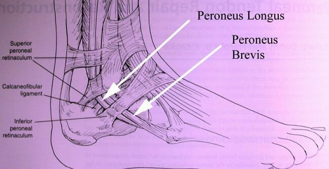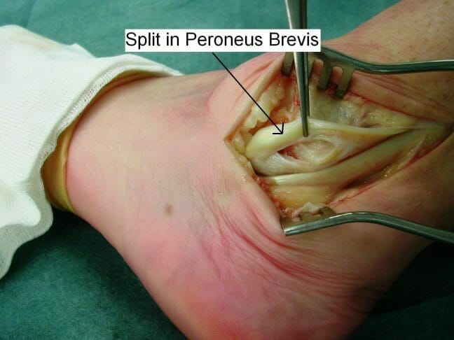

The peroneal tendon runs behind the lateral malleolus (the bony bit on the outside of the ankle). If the tissue that holds the tendons in place is torn by turning the ankle over (ankle sprain) then the tendons can slip forward over the malleolus. The common pathology is tear of the superior peroneal retinaculum and striping of periosteum from the anterior attachment to the lateral maleolus.
The peroneal tendons are held in a grove behind the fibula by the superior peroneal retinaculun.
Subluxation of the peroneal tendons is a rare injury that is often misdiagnosed. This injury is usually related to and often accompanied by lateral ankle ligament sprains. In recurrent subluxation, patients usually give a history of previous ankle injury, which may have been misdiagnosed as a sprain. An unstable ankle that gives way or is associated with a popping or snapping sensation is another common complaint. The peroneal tendons may actually be seen subluxing anteriorly on the distal fibula during ambulation.
Subluxing peroneal subluxation can result from sharp cutting and turning movements. The subluxation can also occur from a direct blow to the outside ankle. The retinaculum, or tissue that encloses the peroneal group ruptures and allows the tendons to snap out of their groove. The athlete will complain of snapping when running or jumping and back again when stress is reduced.
Diagnosis
The diagnosis is usually made clinically by making the patient pull their foot up and out against resistance which will cause the tendon to flick over the ankle. The diagnosis can be confirmed by MRI and more specifically by dynamic ultrasound. The MRI scan will usually show tears in the peroneal tendons which co-exist with this condition. On occasion the tendon which subluxes over the ankle is actually only part of the tendon and represents the torn piece.
The ultrasound scan is performed dynamically by moving the ankle at the same time the probe is scanning. This allows us to actually see the tendon flick over the bone. The video shows a dynamic ultrasound, showing the peroneal tendon (white) flicking over the ankle bone (black).
Make An Enquiry
Or contact us directly
[email protected]
0161 445 4988
